Hook Removal
The guidance on this page focuses on surgical removal in a hospital setting. Dr. Mariluz Parga has created two excellent videos on sea turtle handling and hook removal in a field setting, designed specifically for longline fishermen. These videos are available in ENGLISH and in SPANISH.
 Hooks may snag on soft tissue or become ingested during foraging. There are many types of hooks: single barb, treble, circle and J hooks are used commonly. Refer to "Fish Hook" for descriptions of various hooks. The type of hook, location of attachment (if attached), presence of line or leader, and size of patient will determine the ideal method of extraction. However, resources vary and sometimes salvage procedures are the only option.
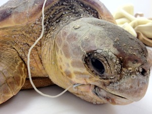
If the hook is visible from the mouth, extraction by way of the oral cavity is advised. If there is line extending down the intestinal tract that is not easily removed with light pressure, cut it as soon as possible to relieve tension. Many oral hooks can be removed safely without anesthesia.
Needle nose or long handled pliers are most effective. The hook should be guided thru the soft tissue in an antegrade fashion. The upright portion of the hook, the shank, should be cut, removing the eye. This will minimize drag and trauma to the tissue as it is pulled through. A scalpel can be used to nick the skin over the point of the barb to facilitate passage through thick dermis.
If the hook is in the esophagus and cranial to the acromion attachment, the best option is esophagostomy. This technique will be described in detail in the Surgical Technique area of our website. Hook removal may also be accomplished with the ARC Dehooker, but significant damage to the esophagus may result. For instructional video on this procedure see ARC Dehooker instructional videos. Additionally, hooks often perforate beyond the esophagus into the cervical region where blind removal via retrograde action is not advised. Damage to vital structures in the area is likely without adequate visualization.
If excessive bleeding is noted after a necessary blind hook removal, a pressure bandage and elevation of the head and neck can be used to allow a clot to form. In an emergency situation where a major vessel has been lacerated, it can be permanently and safely ligated to allow for collateral circulation and compensation.
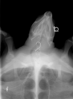
If the hook is beyond the thoracic inlet, follow-up radiographs are needed to see if it is moving. The GI transit time of sea turtles is much longer than in mammals, often requiring weeks for items to pass. If the turtle is eating and passing stool, surgery is not indicated. Surprisingly large hooks can be passed without complications, but follow up radiographs are needed to ensure that the hook is moving steadily abroad. Of far greater concern is mono-filament fishing line that can anchor proximally in the gut and act as a linear foreign body. This will ultimately create an intussusception, perforate the intestinal tract and result in coelomitis.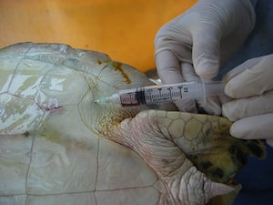 If you are suspicious of a linear foreign body in the gut, it is advised to aspirate the coelom via the inguinal fossa. Large amounts of free fluid tend to accumulate in the coelom when a blockage is present. This blockage may or may not be evident radiographically. This technique is described in detail in the Sample Collection area of our website. Ultrasound is especially helpful to evaluate for the presence of fluid.
Barium studies are of little utility in sea turtles as the GI transit time is already prolonged (often >48 hours) and blockages commonly result in mechanical and or physiologic ileus, which may delay barium passage indefinitely.
If surgery is required to remove a hook located distal to the acromion attachement that is not moving then gastronomy and or enterotomy are indicated. A bilateral pre femoral approach can be used to exteriorize most of the GIT however will cause tension on stomach and intestinal tract to exteriorize them. For large hooks in juvenile and subadult patients that may have the heart imperiled by hook movements an axillary approach is preferred. These approaches and surgical techniques are outlined in Surgical Techniques. A transplastron approach is not advised due to complications healing the incision resulting in lengthy hospitalization. Prompt intervention is the key to success with these cases as the GIT can become quite edematous and friable. The prognosis worsens with the degree of coelomitis present.
If lead sinkers have been swallowed along with the hook, chelation therapy is advised following removal. See Formulary.