Radiographic Technique
Radiographs provide critical information that can greatly affect the outcome of rehabilitation efforts. Many small animal veterinarians and most veterinary hospitals have their own, in house x-ray equipment. If this is not available consider fostering a relationship with a human hospital or clinic that may be willing to help. There is a printable PDF at the bottom of this page that can help orient X-ray technologists to a sea turtle technique.
Xrays are needed for diagnosis of:
Hook ingestion, location, and position
Pneumonia
Buoyancy disorders
Pneumocoelom (free air in coelomic cavity)
Trauma/fracture
Osteomyelitis/arthritis
Obstipation
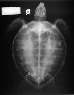 Prior to taking radiographs barnacles are removed from the carapace and plastron. Barnacles will appear on radiographs as bone density tubercules and confuse image interpretation or worse, obscure actual lesions. Care should be taken to remove barnacles on the carapace so as not to damage the scutes. Pressure at the base of the barnacle with a periosteal elevator, screwdriver, or chisel should be sufficient to pry the barnacle loose. Sometimes a gentle tap with a hammer (on the instrument) may be necessary to remove large barnacles.
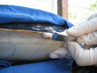
Barnacles on soft tissue can often be removed by picking them off by hand. If the turtle is quite debilitated, it is recommended to remove only the largest and, therefore most radiodense barnacles immediately, saving the rest until the turtle is stronger. If barnacle removal results in damage to scutes or dermis, delay removal until the patient can tolerate removal without injury.
Sea turtles lack a diaphragm that separates thorax from abdomen and instead have a common coelomic cavity. Therefore, horizontal beams must be utilized to evaluate the lateral and anterior-posterior views. Typical placement in right or left lateral recumbency would result in the viscera shifting ventrally and confusing the image. Most x-ray machines can be modified to take horizontal beams by moving the anode tube to the far end of the table, rotating it 90 degrees and placing it ON the tabletop. The focal film distance from machine to plate should be kept at 40 inches (~101 cm) to avoid image magnification. Keep both the beam and patient planes parallel to the table top and perpendicular to the plate to avoid distortion.
Positioning:
1. Dorsoventral or DV. This view is used primarily to evaluate the coelomic cavity. Larger individuals may need to be split onto multiple plates for complete viewing.
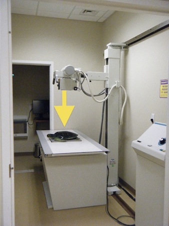
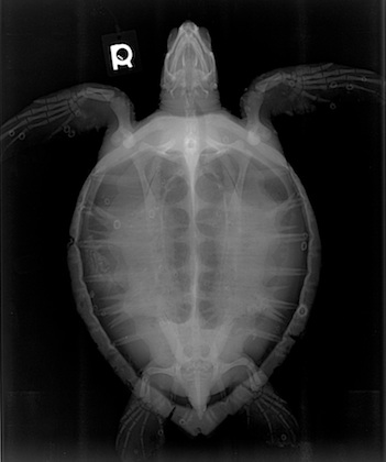
2. Lateral. This right lateral view is used primarily to evaluate cranial and caudal lung fields. Make sure that the turtle is as close to the plate as possible to minimize magnification. Larger individuals may need to be split onto multiple plates.
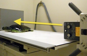
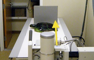
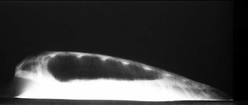
3. Anterior-posterior or AP. This technique is used primarily to evaluate right and left lung fields. It is very important to have the turtle straight for this image, although normal anatomical variation may make perfect positioning impossible.
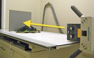
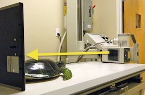

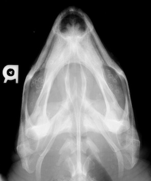
4. Skull DV. This view is mostly used to evaluate for trauma and to ensure that no hooks are present. Many times the skull will be included on the DV film. Although to really appreciate the bone structure, precise measure of the skull and deep bone technique are advised.
Technique:
A “turtle technique” may not be needed to obtain high quality films. Depending on your equipment, either standard abdominal technique or deep bone technique can be used. Measurements should be taken at the highest point of the carapace for the DV view. Lateral technique uses the settings as the DV. AP is done simply doubling the time of the DV (i.e., from 1/30 to 1/15 of a second.)
For skull films, use standard measurements and deep bone technique.
If your equipment does not produce adequate images with the above, the following techniques were suggested by the Turtle Hospital of Marathon and are a place to start for development of a sea turtle technique chart:
Up to 10 kg - 60 Kvp, 12.5 Mas
10-kg kg - 70 Kvp, 20 Mas
Over 50 kg – 100 Kvp, 20 Mas
Resources
The Beauty of Grey: Radiographic technique and positioning in sea turtles PDF
This chapter should be cited: Mettee, Nancy. 2014. Radiographic Technique. Marine Turtle Trauma Response Procedures: A Veterinary Guide. WIDECAST Technical Report No. 17. Accessed online [date].
Updated 3/2014