Physical Exam
All patients should be given a chart (paper or electronic) in which the stranding report, weights, feeding records, treatments, and observations are recorded. Each chart should have a unique identifier to link it to the patient. Charting is of vital importance for future reference and to serve as a record of successes and failures.
Accurate diagnosis requires a minimum database of information. Resources may vary but this is an ideal minimum database:
• History (nature of stranding)
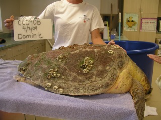
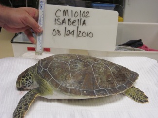
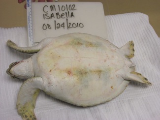
• Digital photos (3 minimum); patient ID and date should be visible in photo
• Weight & measurements
• Complete physical exam with nutritional assessment
• Full body radiographs
• Blood work (i-STAT, CBC, chemistry panel)
Ophthalmic/Otic: Evaluate eyes with natural light and white light. Include fluorescein dye test for corneal ulceration. 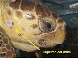
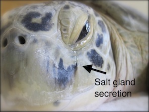
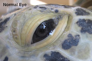
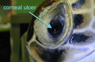
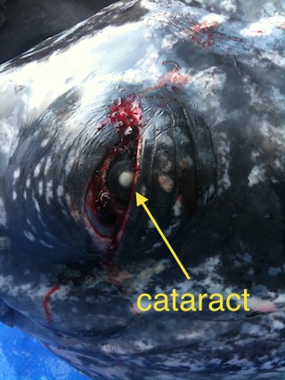
Neurologic: Any individual that displays neurologic signs is given a full neurological exam. This exam is both in the water and out of the water. Serial exams are needed to evaluate progression. Neurological exam form PDF
Musculoskeletal: Carapace and plastron are evaluated for fractures or instability. All limbs should be palpated for range of motion, swelling or crepitus. Evaluate muscle tone and strength. Failure to use a limb in the water constitutes lameness or paresis. Note any skeletal deformities.
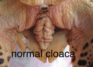
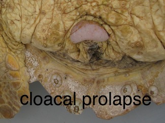
Digestive: The majority of the digestive system is inaccessible as it is between the carapace and the plastron. Examine the oral cavity for foreign bodies or exudate. Perform cloacal palpation. Cloacal prolapse is common in individuals who have been out of the water or are suffering from intestinal impaction/obstruction.
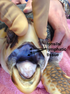
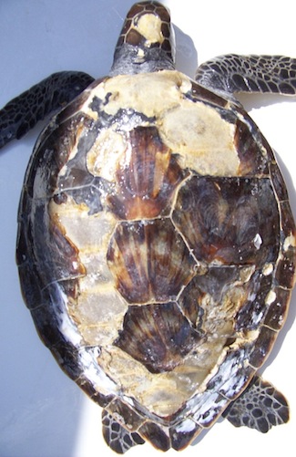
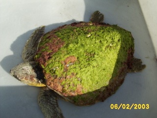
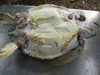
Dermatologic: Scutes cover bone. Many scutes have specific anatomical names. Scales cover skin. Skin should be evaluated for excessive sloughing, wounds, abscesses, and scars. The thickness of a sea turtle's skin makes bruising invisible in most instances. Monofilament should be removed with medical supervision as blood vessels may be constricted in the foreign material and profuse bleeding may occur AFTER removal. Any wound should be assessed by both duration and location. For example, designate acute vs. chronic or soft tissue vs. carapace/plastron. Describe type and distribution of parasites and epibiota.
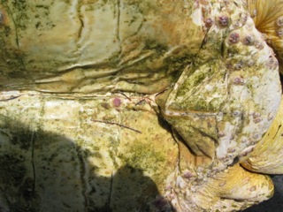
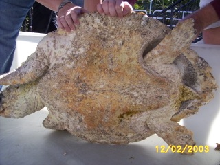
External parasites should be removed. Prior to radiography, barnacles must be scraped off the carapace and plastron. Most barnacles are easily removed, although in extremely debilitated patients their removal may result in loss of the scute leading to bone exposure. Caution should be taken so as not to create further damage. Leeches are removed by soaking the turtle in fresh water for no more than 12 hours. Care should be taken to scrape off the leech eggs as well. Note: if extensive soft tissue damage is present, fresh water will be damaging.
Cardiac: The sea turtle heart has three muscular chambers: two atria and one ventricle. This configuration results in the mixing of oxygenated and non-oxygenated blood, but due to shunting and the overall capacity of sea turtle red blood cells for carrying oxygen, this does not have much impact clinically. It will affect EKG interpretation as a single peak pulse wave will be seen and not a typical PQRS. Peripheral pulses are not palpable and cardiac auscultation is not possible due to the presence of the external shell.
Cardiac evaluation is possible with:
Doppler flow meter with probe placed over a major vessel.
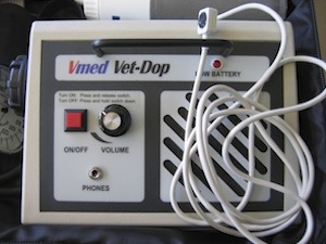
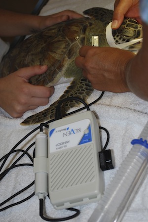
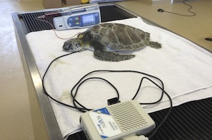
EKG lead II placement (will yield a pulse wave only, not a PQRS). Lead placement close to the body and with metal attachments to patient with skin staples or hypodermic needles can enhance the EKG trace in smaller patients.
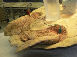
Ultrasound can visualize movement of the heart, left and right aortic arches, and pulmonary arteries using the left and right cervicobrachial area as a window. (need an image)
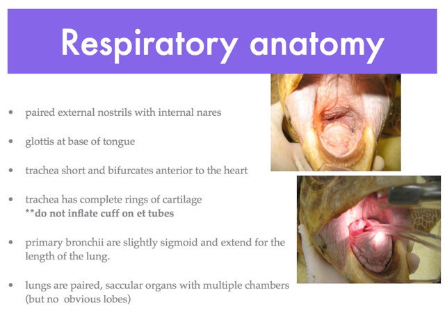
Respiratory: Respiratory rate will vary with temperature and activity level. It is not unusual to see active juveniles breathe every 10 seconds, whereas a debilitated adult may only breathe every 5 minutes or longer.
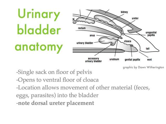
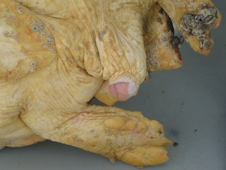
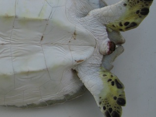
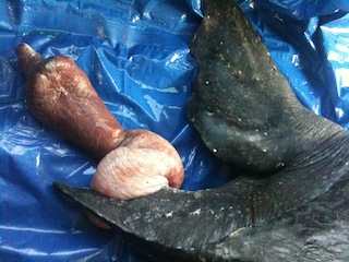
More significant prolapse, such as the rectal prolapse seen below, may require surgical intervention to correct but the underlying cause should be also determined.
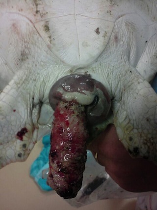
Males may exhibit penile prolapse in addition to cloacal prolapse. It is important to return this tissue to normal position as soon a possible.
Reproductive: Sex should be recorded in the medical record, if known. Sea turtles cannot be externally sexed until adulthood which is determined by carapace size. Males will have a tail that extends beyond the carapace. Female tails will not extend beyond the carapace. Approximate carapace sizes for mature turtles by species are:
> 55 cm Lk,Lo
> 90 cm Cc, Cm
> 80 cm Ei
> 130 cm Dc
(need images of male and female tails)
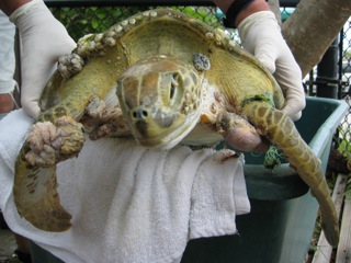
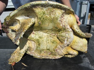
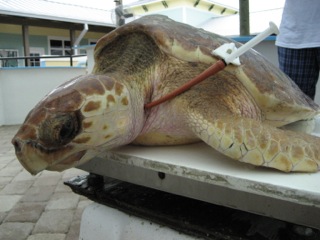
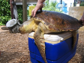
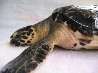
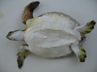
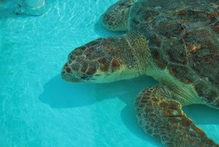
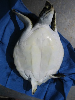
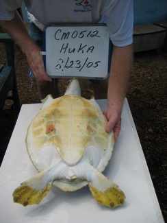
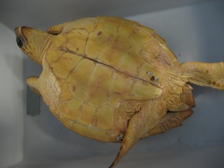
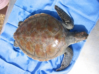
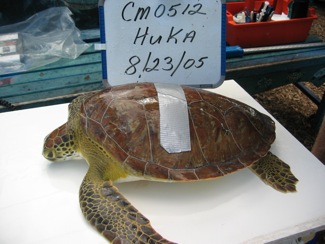
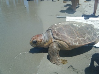
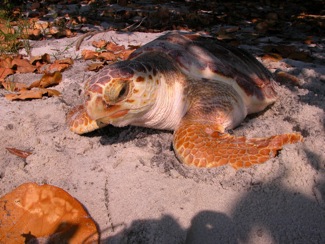
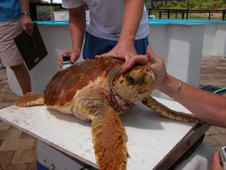
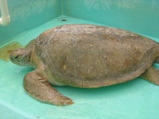
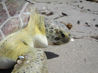
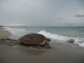
Nutritional status: Evaluate whether the patient is emaciated, thin, adequate, robust.
- Emaciated:
- Sunken eyes
- Loss of shoulder and neck musculature and fat
- Loss of inguinal fat
- Muscle tone watery/loose
- Skeletal elements prominent on skull and plastron
- Frequently loss of soft tissue between bones of carapace and plastron is evident
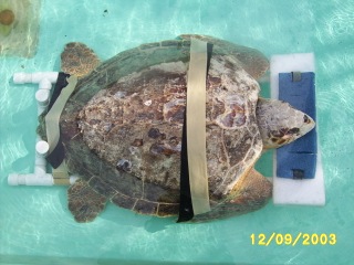
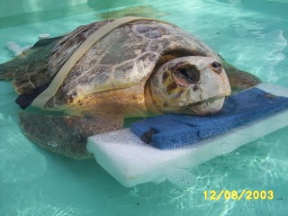
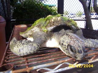
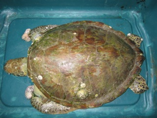
- Thin:
- Loss of fat stores in shoulder, neck, and groin
- Plastron slightly sunken in (concave)
-
Adequate:
-
Normal muscle tone
-
Fat stores present
-
Plastron flat
-
-
Robust:
-
Fat stores present and notable in neck, shoulder, and groin
- Plastron may appear bowed (convex)
-
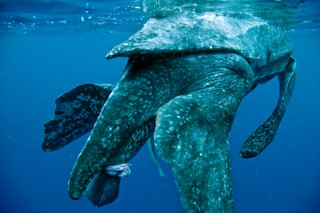
In-water Exam: Turtles are also evaluated in the water for activity levels and behavior. Once a turtle has been placed in the water an adjustment period is common. These are wild animals which have never been confined but are now faced with barriers and walls. This can be stressful and they may enter a panic phase with rapid respiration and hyperactivity/agitation. The panic phase should be short lived (a few minutes); if it is prolonged then removal from the water is warranted.
If the patient is debilitated, observation for 5-10 minutes is needed to ensure that the patient can get its head out of the water to breathe and that breaths are regular. Gurgling or weak respirations warrant further dry dock or support in the water. Caution must be used when patients are on support floats in the water and supervision is required for the safety of the animal.
Any buoyancy abnormality should be recorded with anatomic location (right cranial, central caudal, etc). It is also helpful to estimate the percentage of carapace exposed. This will allow for serial examination. If the air/gas is moving it is more likely intestinal gas and less likely in the lungs. If the buoyancy is resolving then intervention may not be required.
For further discussion of buoyancy disorders see Buoyancy Disorders
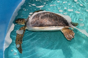
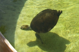
"Entire left side, 40% carapace exposure" "100% submerged, rear buoyant"
Radiographs: All cases are radiographed with three standard views. These are detailed in the Radiographic Technique section.
Dry Dock: Incoming patients are all “dry docked” (out of the water) for the first night to avoid drowning due to weakness or hypoglycemia. Capture myopathy is a sudden weakness due to massive muscle exertion and often will develop hours after capture as the adrenaline effect subsides. Even apparently calm turtles should be dry docked as stress levels are very high for these wild animals. Dry docked individuals should be place in a bin for confinement.
After assessment in the water the following day, turtles that have questionable strength are further dry docked until they demonstrate sufficient strength so that drowning is no longer a concern. Sea turtles can tolerate extended periods of time out of the water, even weeks in some instances, if certain precautions are taken. Circulatory impairment out of the water is a major concern. Signs of right-sided heart failure (ascites, limb edema) may become apparent early in large individuals, and cloacal prolapse is common. To avoid these more common issues, very weak turtles can be put on flotation devices and placed in the water for hours each day. This will improve circulation to the extremities, but it requires supervision to ensure that the turtle does not fall off the float.
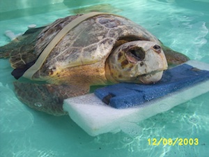
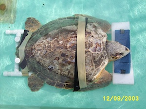
Dry docked individuals should have the skin coated in an occlusive ointment such as Vaseline or A&D ointment to prevent drying and cracking. The eyes will need to be lubricated with an ointment twice daily (do not use drops since they will evaporate more quickly requiring more frequent application). Cushions and pads should be placed beneath the turtle to prevent decubital ulcer formation (pressure sores). These pads will need to be changed and/or cleaned regularly.
This chapter should be cited: Mettee, Nancy. 2014. Physical Exam. Marine Turtle Trauma Response Procedures: A Veterinary Guide. WIDECAST Technical Report No. 17. Accessed online [date].
Updated 6/2013
